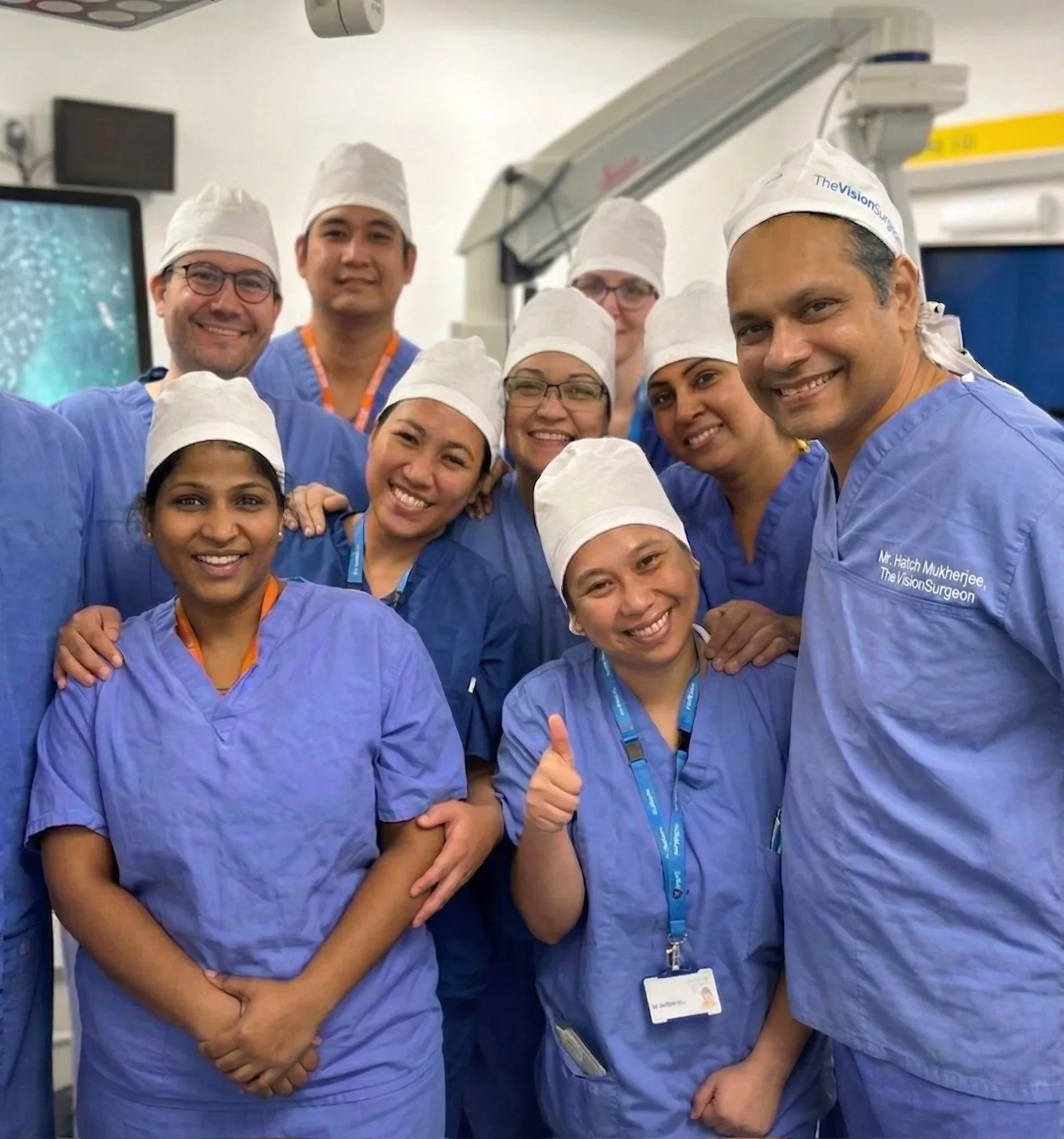
MIGS Fellowship · King’s College Hospital · FRCOphth · FWCRS · Colchester, Essex
Glaucoma Treatment in Essex
Specialist glaucoma care led personally by Mr Hatch Mukherjee — fellowship-trained in minimally invasive glaucoma surgery (MIGS) at King’s College Hospital London. From early SLT laser through to iStent and advanced surgical options, your care is consultant-led from diagnosis to long-term management.
Mr Hatch Mukherjee is one of the first surgeons in the UK to perform both CAIRS and topography-guided laser surgery for keratoconus — bringing techniques previously available only in specialist centres to patients across Essex and East Anglia.
Professional memberships, fellowships & leadership roles


What is Glaucoma?
Glaucoma refers to a group of eye conditions that damage the optic nerve — the nerve responsible for carrying visual information from the eye to the brain. In most cases this damage is linked to raised pressure inside the eye, though glaucoma can also develop at normal pressure levels.
It is often called the ‘silent thief of sight’ because it typically causes no noticeable symptoms until significant damage has already occurred. By the time peripheral vision is affected, the condition has usually been present for some time. This is why regular eye examinations are essential, particularly for those with risk factors.
Although glaucoma cannot be reversed, it can be effectively managed. With early detection and the right treatment, most patients retain good vision throughout their lives

Types of glaucoma
Recognising the Different Forms
Glaucoma is not a single disease. Understanding which type you have determines the most appropriate treatment pathway.
Open-Angle Glaucoma
The most common form. The drainage angle between the iris and cornea remains open, but the drainage tissue (trabecular meshwork) becomes less efficient over time, slowly raising eye pressure and damaging the optic nerve. Often asymptomatic until advanced.
Angle-Closure Glaucoma
The drainage angle narrows or closes suddenly, causing a rapid rise in pressure. An acute attack is a medical emergency — symptoms include sudden eye pain, headache, blurred vision, halos around lights and nausea. Requires urgent treatment.
Normal-Tension Glaucoma
Optic nerve damage occurs despite eye pressure being within the normal range. Reduced blood supply to the optic nerve is thought to play a role. Careful monitoring and pressure-lowering treatment remain the mainstay of management.
Secondary Glaucoma
Raised pressure resulting from another eye condition, injury, inflammation, or long-term steroid use. The underlying cause is treated alongside pressure management. Common examples include pseudoexfoliation and pigmentary glaucoma.
RISK FACTORS
Who is at Risk?
Glaucoma can affect anyone, but certain factors significantly increase the risk. Knowing your risk profile is the first step towards protecting your sight.
- Family history — a first-degree relative with glaucoma significantly increases your risk
- Age over 40 — risk increases progressively with age
- Raised intraocular pressure — the primary modifiable risk factor
- Ethnic background — higher prevalence in African, Caribbean and Asian populations
- High myopia — short-sightedness increases susceptibility to certain glaucoma types
- Long-term steroid use — particularly steroid eye drops can raise eye pressure
- Diabetes or cardiovascular disease — associated with optic nerve vulnerability
**Urgent assessment is needed: **if you experience sudden eye pain, blurred vision, halos around lights, or nausea — these may indicate an acute angle-closure attack.
DIAGNOSIS
How Glaucoma is Diagnosed
Accurate diagnosis requires a thorough assessment using specialist equipment. At The Vision Surgeons, all investigations are personally overseen by Mr Mukherjee.
Tonometry
Measures intraocular pressure. Raised pressure is a factor, though pressure alone does not confirm or exclude glaucoma.
Visual Field Testing
Computerised perimetry maps your peripheral and central visual field to detect areas of vision loss caused by optic nerve damage.
OCT Scanning
Optical coherence tomography provides detailed imaging of the optic nerve and retinal nerve fibres for detecting damage before visual field changes appear.
Gonioscopy
Examination of the drainage angle to determine whether it is open, narrow, or closed — essential for classifying the type of glaucoma and guiding treatment.
Treatment options
A Stepwise Approach to Glaucoma Management
Treatment is tailored to the type and severity of glaucoma, your response to previous therapy, and your overall eye health. Mr Mukherjee offers the full range of options — from first-line laser treatment through to surgical intervention.
Eye Drops
Pressure-lowering eye drops remain the most commonly used treatment for early glaucoma. Several classes are available, each working through different mechanisms — either reducing fluid production within the eye or improving drainage. The right drops depend on your pressure target, lifestyle and tolerability.
- Prostaglandin analogues — once daily, highly effective
- Beta-blockers — reduce fluid production
- Carbonic anhydrase inhibitors — oral or topical
- Combination drops — simplify regimen when multiple agents are needed
Selective Laser Trabeculoplasty (SLT)
SLT is a non-invasive outpatient laser procedure that targets the drainage tissue (trabecular meshwork) inside the eye. Short, low-energy laser pulses stimulate the body’s natural drainage mechanism, improving fluid outflow and reducing eye pressure.
- No incisions — performed with anaesthetic drops only
- Takes approximately 10–15 minutes
- Can be used as a first-line treatment or alongside drops
- Can be repeated if the effect diminishes over time
- Evidence supports SLT as effective as drops for many patients (LiGHT trial)
Minimally Invasive Glaucoma Surgery (MIGS)
MIGS procedures are designed to lower intraocular pressure with less tissue disruption and faster recovery than traditional glaucoma surgery. Mr Mukherjee completed his fellowship in MIGS at King’s College Hospital London — one of the UK’s leading centres for surgical glaucoma management.
iStent — a microscopic titanium implant placed within the eye’s drainage system, often combined with cataract surgery. It acts as a permanent bypass, allowing fluid to drain more freely and reducing pressure with minimal disruption to the eye’s anatomy.
GATT (Gonioscopy-Assisted Transluminal Trabeculotomy) — a suture-based procedure that opens the drainage canal (Schlemm’s canal) through a very small incision, lowering resistance to fluid outflow across the entire drainage system.
Trabeculectomy & Tube Shunts
When drops, laser, and MIGS are insufficient to control pressure adequately, conventional filtration surgery may be recommended. These procedures create new drainage pathways to achieve a sustained and reliable reduction in eye pressure.
Trabeculectomy creates a small drainage bleb under the conjunctiva, allowing aqueous fluid to filter out of the eye at a controlled rate. It remains one of the most effective procedures for significant pressure reduction.
Preserflo MicroShunt — a small implantable drainage device that creates a controlled pathway for fluid to leave the eye, with a lower risk of complications compared to traditional tube shunts and a faster visual recovery.
Cross-linking, CAIRS, topography-guided laser — the full range of advanced corneal treatments in one place.
Book a consultation — discover The Vision Surgeon AdvantageSame consultant from first assessment through to treatment and follow-up
Why Choose Mr Hatch Mukherjee?
Mr Mukherjee brings specialist fellowship training in glaucoma and MIGS alongside his expertise in refractive surgery — a combination that is rare outside major London centres. Patients with glaucoma who are also considering or undergoing cataract or lens surgery benefit directly from this dual expertise.
MIGS Fellowship — King’s College Hospital, London
Fellowship-trained in minimally invasive glaucoma surgery at one of the UK’s most respected centres, working alongside internationally recognised glaucoma specialists.
Combined Cataract & Glaucoma Expertise
Many patients with glaucoma also develop cataracts. Mr Mukherjee’s expertise in both allows combined procedures that address both conditions in a single operation — reducing risk and recovery time.
Advanced Diagnostic Technology
OCT imaging, visual field testing and tonometry are all available on-site, providing the detailed information needed to detect glaucoma early and monitor progression precisely.
You See the Consultant at Every Visit
All assessments, treatment decisions and follow-up appointments are led personally by Mr Mukherjee. Long-term glaucoma management demands consistency — the same experienced eye sees your optic nerve every time.

What to Expect at Your Consultation
- Initial consultation — Mr Mukherjee reviews your history, performs a full slit-lamp examination and reviews any previous investigations. Eye pressure is measured and the optic nerve assessed directly.
- Imaging and investigations — OCT scanning of the optic nerve and retinal nerve fibre layer, corneal pachymetry (thickness), gonioscopy (drainage angle assessment), and visual field testing where appropriate.
- Diagnosis and classification — findings are discussed clearly and honestly. Where the diagnosis is uncertain, further monitoring may be recommended before committing to treatment.
- Treatment planning — if treatment is needed, Mr Mukherjee explains each option, its evidence base, likely effectiveness for your specific case, and what to expect from recovery. You decide together.
- Long-term monitoring — glaucoma is a lifelong condition. Regular review appointments track pressure, optic nerve appearance and visual field to ensure the treatment target is being met and to detect any progression early.
Frequently Asked Questions
RELATED TREATMENTS
Not sure laser is right for you?
If laser eye surgery is not suitable — due to a high prescription, thin corneas or other factors — there are excellent alternatives that Mr Mukherjee can discuss at consultation.
Laser Eye Surgery
Once keratoconus is stabilised, selected patients may be candidates for topography-guided laser correction to reduce residual refractive error.
Lens Replacement Surgery
For patients with keratoconus and presbyopia, lens replacement with a premium IOL can address both conditions simultaneously.
Cataract Surgery
Keratoconus patients developing cataracts require particularly careful biometry and lens selection — an area of specialist expertise for Mr Mukherjee.
Specialist glaucoma care in Colchester, Essex.
Mr Hatch Mukherjee provides fellowship-trained diagnosis and management of glaucoma — from first-line laser treatment to advanced MIGS and surgical options. All care is personally led by the consultant throughout.